


In brain tumor surgery, both the extent of resection and the timing of the operation influence the outcome. The decision about whether to operate, when, and by what approach is therefore taken carefully, on the basis of imaging, tumor type, and the patient’s overall condition.
Artificial intelligence as an assistant in decision-making
The Personalized Evidence-Based Treatment Analysis uses AI to compile, for each patient, a structured review of the current literature as it applies to their specific characteristics — age, imaging findings, comorbidities, prior treatment. Dr. Blionas reviews this report and uses it to test and refine his treatment recommendation. The result is a decision grounded in evidence that would otherwise take weeks to assemble.
Each patient also receives a written summary of their history and condition, setting out the options with their advantages and drawbacks, which they are free to take to another doctor for a second opinion.
The aim of surgery
The goal is maximal safe resection: removing as much of the tumor as can be removed without putting the patient’s function at risk. These two considerations — how much is removed, and what it costs the patient — are weighed against each other in every case.
The extent of resection matters. In benign tumors, complete removal substantially reduces the chance of the tumor returning compared with partial removal. In high-grade malignant tumors, the published evidence consistently links a greater extent of resection with longer survival, and removing the great majority of the tumor appears to confer a benefit over biopsy alone.
Equally important is that the removal is carried out safely, without injury to the eloquent areas of the brain — the regions that control movement, sensation, speech, and thinking. A more complete resection that leaves the patient with a permanent deficit is not a better result. Where the tumor lies within or immediately adjacent to these areas, a deliberately incomplete resection is sometimes the correct decision.
How the operation is performed
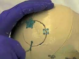
Surgery for a brain tumor involves a craniotomy — the temporary removal of a section of the skull to give access to the brain beneath. The skin is opened and the muscle displaced, the bone is lifted, and at the end of the operation it is replaced and secured to the surrounding skull with small fixation plates. Occasionally, if the brain is swollen at the end of the procedure, the bone is left out and replaced at a second, smaller operation once the swelling has settled.
Beneath the bone lies the dura mater, the tough membrane covering the brain. Once this is opened, the approach depends on where the tumor sits. Tumors on the surface, and those arising from the coverings of the brain rather than within it, are reached directly. For tumors lying deeper, a small opening is made in the surface of the brain to reach them.
The removal itself is carried out with the support of several technologies, each addressing a different problem:
Neuronavigation works like a GPS for the brain. Using the preoperative MRI, it shows the surgeon the position of the instruments in relation to the tumor at any moment.
Intraoperative neuromonitoring is used when the tumor lies near the pathways controlling movement and sensation. These are monitored continuously during the operation, so that any change is detected while it is still reversible.

Brain mapping is a more precise form of the same principle: small electrodes and brief electrical stimulation are used to identify exactly where the functional areas lie, so that they can be avoided.
Awake craniotomy is used mainly when the tumor is close to the speech areas. The patient is woken for part of the operation and asked to speak, name objects, or read while the resection proceeds, so that any effect on language is detected immediately. Patients are prepared for this beforehand, and the part of the operation involving the skull is not felt.
Frozen section gives a preliminary answer from the laboratory while the patient is still in theatre, helping the surgeon judge how far to extend the resection. The definitive diagnosis, which now includes molecular testing, takes longer and follows in the days after surgery.
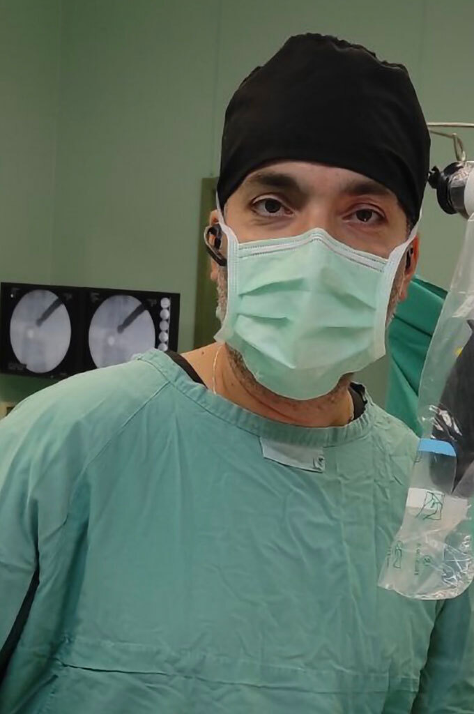
Technically, the resection is performed under a high-magnification operating microscope, or with a 3D 4K exoscope, using microsurgical instruments and working from the periphery of the tumor inwards. An ultrasonic aspirator is often used as well: it fragments and removes tumor tissue with minimal traction on the surrounding brain.
Once the tumor — or the part of it that can be safely removed — has been taken out and sent for analysis, any bleeding is controlled, the dura is closed, and the bone, muscle, and skin are repositioned.

The image shows the tumor centrally, with the surrounding dura. Careful closure of the dura after removal is an important step.
Complications
Every operation on the brain carries risk, and these are discussed in full before any decision is made.
Bleeding within the skull is the most serious early complication; it is uncommon, but in a small number of cases it requires a second operation. Infection and cerebrospinal fluid leak are the relatively more frequent problems. A CSF leak shows itself as a soft swelling at the wound and usually settles with wound care alone; only rarely is further surgery needed to repair the dura.
Seizures can occur after surgery, and preventive medication is sometimes given for a period afterwards. Blood clots in the legs or lungs are a recognized risk after any major operation, and measures are taken from the outset to reduce it.
The complication of greatest concern — and the one that the technologies described above are designed to prevent — is injury to eloquent brain, which can cause weakness, loss of sensation, or difficulty with speech and comprehension, depending on where the tumor lies. Many such deficits are temporary and recover over weeks, but some are permanent.
Overall complication rates depend heavily on the position of the tumor: in the region of 5% for a tumor in a favorable location, and 10% or higher for one in a difficult one.
See also: Transsphenoidal surgery for pituitary and skull base tumors