



Hydrocephalus is a serious condition that can be life-threatening if not treated promptly.
The treatment options are:
- Shunt insertion — ventriculoperitoneal, ventriculoatrial, or ventriculopleural
- Lumboperitoneal shunt
- Endoscopic third ventriculostomy (ETV) — a minimally invasive alternative
On the available evidence, these achieve broadly similar success rates of around 70–80% in appropriately selected patients, and each carries its own advantages and disadvantages.

Careful, individualized selection. In a condition that requires prompt decisions, every option is assessed individually through the AI-supported Personalized Evidence-Based Treatment Analysis, which draws together the current literature and the patient’s own characteristics. The surgeon reviews that analysis to substantiate and refine his choice. Each patient also receives a complete account of their history and condition, with the advantages and disadvantages of every option, which they can present to another doctor if they wish to seek a second opinion. An online assessment is available for patients in remote areas.
Shunt insertion
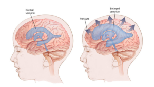
A shunt is a valve mechanism connected by tubing to the cerebral ventricles, which drains fluid whenever the pressure rises above a threshold set by the neurosurgeon for that patient. The fluid is directed through a second tube to a cavity where it can be absorbed — usually the peritoneal cavity in the abdomen, less often the right atrium of the heart or the pleural cavity.
Advantages. The system can be adjusted at will, altering the opening pressure to suit the patient, and the treatment is reversible: the shunt can be set to a non-draining setting or removed altogether.
Overdrainage must be recognized and corrected by adjusting the valve; left untreated it can cause headache and vomiting, and in some cases subdural collections or haematomas.
Disadvantages. A shunt is an implanted foreign body, which may cause discomfort and, more importantly, can become infected — often requiring removal of the system. As a hydraulic system, it is also subject to mechanical failure: tubing may obstruct without warning, and the valve mechanism may fail. Each failure requires further surgery to replace the affected component.
Cumulative failure rates over a patient’s lifetime are substantial, and some patients experience repeated malfunctions. A further consideration is that the proximal part of the operation involves the brain, with the attendant possibility of intracranial complications — infection, haemorrhage, neurological deficit, or seizures — which are less frequent with lumboperitoneal shunting.

The ventriculoperitoneal shunt is the commonest system used for hydrocephalus: a valve drains fluid from the ventricles when the pressure exceeds a set threshold and directs it to the peritoneal cavity.
Lumboperitoneal Shunt
A lumboperitoneal shunt uses the same principle, but the valve is placed in the lower back and the proximal tube draws fluid from the lumbar subarachnoid space. A second tube directs it to the peritoneal cavity. Because the spinal and cranial CSF form a single hydraulic system, lowering the pressure in the lumbar spine lowers it in the head correspondingly.
Advantage. It bypasses the brain entirely, avoiding the complications of intracranial surgery. Where infection or other complications occur, they involve the spine and abdomen rather than the brain, which reduces their overall severity. The valve can likewise be adjusted or closed.
Disadvantages. Failure rates are comparable to, or slightly higher than, those of a ventricular shunt. The system is also less reliable at maintaining stable intracranial pressure, because its function depends significantly on posture: it drains chiefly when the patient is upright and mobile, and less effectively when lying down.
Important: a lumboperitoneal shunt is suitable only in communicating hydrocephalus. Where there is obstruction to CSF flow, lumbar drainage risks downward herniation, and the option is contraindicated. Long-term use may also lead to acquired tonsillar descent — meaning movement of important neural structures with possible complications.

The lumboperitoneal shunt draws fluid from the spine through a small tube and directs it to the peritoneal cavity when the pressure exceeds the set threshold.
Endoscopic Third Ventriculostomy (ETV)
ETV is a minimally invasive procedure in which an opening is created in the floor of the third ventricle, allowing CSF to pass directly from the ventricles to the subarachnoid space around the brain, where it is absorbed. It is, in effect, a surgically created internal bypass — with no implanted hardware at all.
The procedure is performed endoscopically, through a small skin incision and a small burr hole in the skull.
Advantages. No foreign body means no risk of shunt infection, no valve to fail, and a considerably lower likelihood of reoperation than with a shunt. Overall complication rates are in the region of 5–10%.
Disadvantages. There is no control over the rate of CSF drainage, and no way to reduce the function of a stoma that drains excessively — although symptomatic overdrainage after ETV is rare. Being an intracranial procedure, it carries the risks common to brain surgery; and because the stoma is created close to the basilar artery, there is a small (in the order of 0.2%) but potentially catastrophic risk of vascular injury.
Patient selection is decisive. ETV is most effective where there is obstruction to CSF flow — demonstrated on CSF flow MRI, or caused by a tumor — and where the ventricular anatomy is favorable. In well-selected patients its efficacy matches that of a shunt (70–80%, and higher in ideal cases), while its complication profile is better, which makes it the preferable option where the criteria are met. In poorly selected patients, efficacy falls substantially — in some series below 50%. Age and the underlying cause also influence the likelihood of success, and are taken into account.
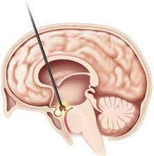
In third ventriculostomy an opening is created in the floor of the third ventricle. Fluid passes through it to the surface of the brain for faster absorption, reducing pressure and symptoms. The image also shows the basilar artery, the principal risk of the procedure.
In summary
Where the criteria are met, ETV is the preferable option, since it achieves comparable results without an implanted system. Where they are not, a shunt is the better choice — ventriculoperitoneal or lumboperitoneal, depending on the type of hydrocephalus and the individual patient, since the two are broadly comparable in efficacy.