



MINIMALLY INVASIVE SPINAL DECOMPRESSION THROUGH A 1 CM INCISION
Treatment of sciatica due to spinal stenosis using minimally invasive lumbar decompression — without general anesthesia, with same-day discharge
A 68-year-old patient presented with lumbar spinal stenosis.
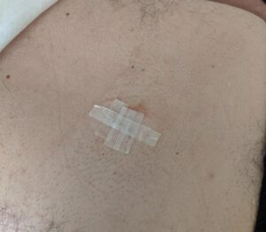
Using a minimally invasive technique, complete decompression was achieved on both the right and left sides through a single incision of approximately one centimeter on the left.
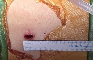
Through this single 1 cm left-sided incision, we gain access to the entire level of the spine — both right and left — decompressing both the right and left nerve roots with minimal bone removal.


Using specialized equipment (tubular retractors), we can focus precisely on the site of the problem and correct it without causing extensive damage to the patient’s body.
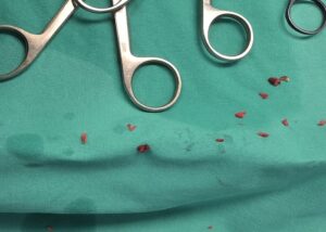
Ultimately, we removed the bony structures that were compressing the patient’s nerves on both sides (right and left).
The patient underwent surgery without general anesthesia and was discharged from the hospital the same day, significantly improved.
Case 1 — Epidural (Transforaminal) Injection
A 50-year-old patient with a lumbar disc herniation compressing the left L4 nerve root. Using a fine needle under fluoroscopic guidance, the injection is delivered precisely along the course of the affected nerve.
The procedure is performed in a state-of-the-art angiography suite, allowing real-time imaging control.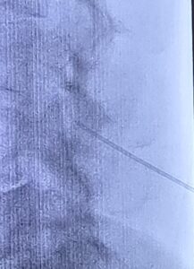
The patient returned to normal activities immediately and has remained symptom-free for months — a safe, effective option for appropriately selected patients.
Case 2 — Awake Same-day Minimally Invasive Microdiscectomy
A 50-year-old patient with a lumbar disc herniation, presenting with severe low back pain and foot weakness.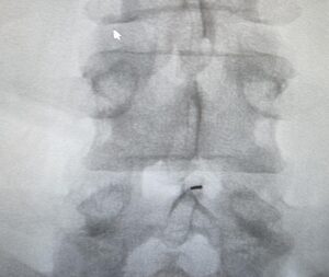
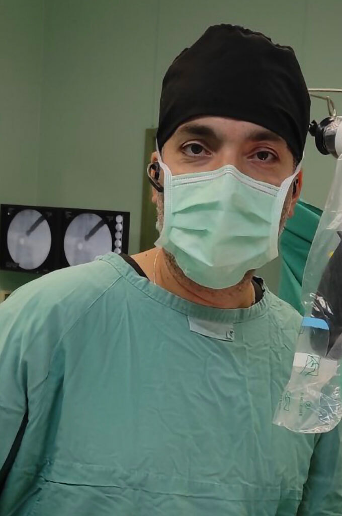
Using the latest technology, we performed a minimally invasive microdiscetomy without general anesthesia: the patient remains awake in the operating room, under sedation combined with epidural anesthesia, in close coordination with the anesthesiologist, while the surgeon proceeds with the microdiscetomy.
Throughout the procedure, three imaging sources are monitored simultaneously: the point of nerve compression on the navigation screen, the fluoroscopic image confirming the exact anatomical level, and the magnified view through the operating microscope.
Finally, the herniated disc material — the primary cause of the patient’s symptoms — was removed. The final incision measures approximately 1 centimeter.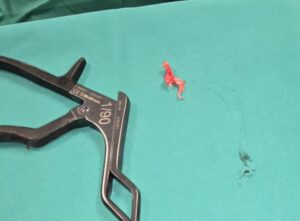
The small incision offers both cosmetic and medical benefits, with a reduced risk of complications such as infection and bleeding.