


Brain and spine procedures require advanced technology in the operating room as a prerequisite for significantly better outcomes with the fewest possible complications. Depending on the case, the equipment used by Dr. Alexandros Blionas includes:
Exoscope (3D 4K video-robotic microscope)
This represents the cutting edge of modern neurosurgery. It replaces the traditional microscope — through which the surgeon looks via eyepieces — with a digital system of extremely high-resolution (4K) cameras that project the surgical field onto large screens. The three-dimensional imaging gives the surgeon a true sense of depth, essential for delicate movements in sensitive anatomical areas such as those near nerves or blood vessels. It also dramatically reduces surgeon fatigue, and because the image is displayed on screens, the entire surgical team sees exactly what the surgeon sees, in 3D — improving coordination and surgical safety.
C-arm Fluoroscopy
Unlike a standard X-ray, which produces static images, the C-arm works like live video: it shows the surgeon the movement of anatomy and instruments continuously and in real time — critical for the dynamic nature of surgery, and without the need to reposition the patient.

It is used to place screws and rods with high precision, and for targeted injections into the spinal nerves for the management of chronic pain. It is essential for procedures that avoid large incisions, since the surgeon can follow the trajectory of each instrument on the fluoroscopic images displayed beside the operating table — reducing the risk of error or injury to adjacent tissues.
Neuronavigation
An advanced computer-assisted system that functions, in effect, as a GPS for the brain and spine. It allows the surgeon to see the precise position of their instruments within the patient’s body relative to the anatomy. Before surgery, the patient undergoes an MRI or CT scan; these images are transferred to the neuronavigation system. Once the patient is positioned on the operating table, the system synchronizes the digital images with the patient’s actual anatomy — typically using specialized sensors or by registering reference points on the skull or spine.
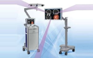
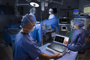
During the procedure, specialized cameras track the surgical instruments, and the surgeon sees their exact position overlaid on the patient’s scans in real time. Neuronavigation helps avoid sensitive areas and critical vessels or nerve pathways, and makes significantly smaller incisions possible.
Intraoperative Neuromonitoring
A technique that monitors the function of the nervous system — brain, spinal cord, and nerves — in real time throughout surgery. Specialized electrodes are placed on the patient (for example on the limbs, face, or scalp); the system delivers small, painless electrical stimuli and records the signals returned by the muscles or nervous system.
If the surgeon approaches too close to a nerve, or applies pressure that could cause harm, the system acts as an alarm — issuing an immediate audible or visual alert. This allows the surgeon to stop or change course before any irreversible damage, such as paralysis or loss of sensation, can occur.
“`